Monday, November 19, 2012

Bibliography


McFarland, Kenneth.Botany 111 Fall 2012 Blog .http://botany1112012.blogspot.com/ .October 21, 2012

Cook R. and McFarland K. General Botany 111 Laboratory Manual.2012.4th ed. 155-157 p.

Botany 111 fall 2012 [internet]. blog spot[cited 2012 october 19] available from      http://botany1112012.blogspot.com/

Patterson D.J. 1998. Free-Living Freshwater Protozoa. New York (NY): John Wiley & Sons Inc.

Smith, Douglas Grant.2001.Pennak’s Freshwater Invertebrates of the United States: Porifera to Crustcea.4th   ed. New York(NY): John Wiley&Sons,Inc.

Pennak, Robert W.1989.Pennak’s Freshwater Invertebrates of the United States: Protozoa to Mollusca.3rd ed. New York(NY): John Wiley&Sons,Inc.

Stremberger Richards. 1979. A guide to Rotifers of the Laurentian Great Lakes. Cincinnati (OH): Academic Press. p. 126-127. (cited 2012 October 25)

Prescott GW. 1964. The Freshwater Algae. WM.C. Brown Company Publishers. 231 p.

Donner J. 1956. Rotifers. Frederick Warne and Company Ltd. 61 p

Micro Aquarium Week Four

FOOD PELLET INFORMATION:"Atison's Betta Food" made by Ocean Nutrition, Aqua Pet Americas, 3528 West 500 South, Salt Lake City, UT 84104. Ingredients: Fish meal, wheat flower, soy meal, krill meal, minerals, vitamins and preservatives. Analysis: Crude Protein 36%; Crude fat 4.5%; Crude Fiber 3.5%; Moisture 8% and Ash 15%.(McFarland)

This week, on November 15th I observed three new organisms in my aquarium. Almost every organism I observed last week has flagella, making it a moving organism. Most were very fast, and others were pretty slow. 

The first one I came across was the Cyclidium sp. He was not as fast as the others, but still on the go.
As one can easily conclude, this organism has flagella surrounding its entire body. It also has a tail flagella which is difficult to see in this picture as it is very poor quality. Its body is ovular, as you can see, and is multi-cellular. One can also observe that this organism contains chlorophyll as the green pigment hints. This organism tends to stay in the middle-range of my aquarium; I did not find any in any other location. This is the only organism of its kind that I observed in my aquarium. 

The next organism I found was a Euchlanis sp. This was the most interesting to me as the body of this organism is very unique.
The first thing one notices is the one flagella located at the rear of the organism. This accounts for why it is not a very fast organism. I had no trouble keeping up with this little guy. He was also very photogenic as this is the best picture I got this week. He also seems to have  a green pigment on him which suggests he contains chlorophyll. The way this organism is easily identified is its unique body shape. It has a body that looks like two balloons being pushed together with a tail hanging off and a mouth on the opposite end. This organism was not very common in my observation of the micro aquarium. This was the only one I observed of its kind.

The third organism I encountered was an Actinophrys sp. 

As one can observe from the images above, the Actinophrys sp. is round in shape and just like the Euchlanis, it has flagella surrounding its body to propel it and give it mobility. This organism is very fast and actually has a very interesting and different mode of transportation. As it is propelled, the shape of the organisms quickly changes to a star-shaped structure; you can almost see this forming in the first picture above. This organism also appears to contain chlorophyll due to its green pigmentation. This organism is very abundant in my aquarium. In fact, on a much lower lens, you can observe many of them at a single time as little dots swimming around. 

The other organisms previously seen in my tank are still present, however not much progress or change has occurred among many of those species. I observed the cyclops sitting still this week and feeding, not moving or anything, and he did not seem any bigger than the last time I observed him. 
The only other observation I made was that the snail population had greatly decreased. I believe this is due to my accident the week previous to this one, in which I spilled out much of the material and water in my aquarium. I am not sure if the organisms found this week came from the changes in the aquarium, or if I had just missed them in previous lab observations.
And, lastly, the smaller Annelid I had observed last week has grown (although not as much as the larger one), and is now feeding as the other one did. However, the larger Annelid I previously observed seems to have died and decomposed. 

It has been very interesting to see how things have changed in just four weeks in such a small habitat. 

So, this week I observed:
- 3 new organisms~
1) Cyclidium
2) Euchlanis
3) Actinophrys
- Snail population decrease
- Decrease in organism movement
- Dead and bigger Annelids

Monday, November 12, 2012

Micro Aquarium Week Four

This week I viewed my aquarium on Thursday, November the 8th at 11 am. To observe it I used a compound microscope in the lab and a Sony camera to take pictures with. I also used Photoshop to edit the pictures for the purposes of this blog.

FOOD PELLET INFORMATION:"Atison's Betta Food" made by Ocean Nutrition, Aqua Pet Americas, 3528 West 500 South, Salt Lake City, UT 84104. Ingredients: Fish meal, wheat flower, soy meal, krill meal, minerals, vitamins and preservatives. Analysis: Crude Protein 36%; Crude fat 4.5%; Crude Fiber 3.5%; Moisture 8% and Ash 15%.(McFarland)

This week I observed what was, in my opinion, the most interesting species so far: a microscopic Annelid (Smith 2001). It was nestled inside the dirt at the bottom of the tank feeding, so I did not get many clear pictures of him, but I did get a video (shown at the bottom of this post).
It can be observed that it is an annelid as it has microscopic hair on its body, which is characteristic of this species. Its movement was mostly in a straight line, unless it was turning, then it would curve over itself to keep going. This was no small organism, however. This annelid was at least 3-4 lens views long, and kept me adjusting. It was interesting for me, to see how large this organism is in comparison to the others in my tank.

The next thing I observed was the cyclops Copepoda from last week (Pennak 1989). It appears to have grown slightly, and I was able to get a better, more clear picture of it.

This week I did not find a single midge, which seems very odd in comparison to a week ago. This could be due to the fact that halfway through my observations, when taking the aquarium off of the microscope plate, I tipped the aquarium too much sideways, and a lot of water was lost in spillage. However, the observations I had made prior to the spill indicate no midge present. It is also important to mention that the long worm I observed last week was also not present.

On a brighter note, I found a new organism!
It is also called a annelid, but looks significantly different from the one above. This one has clear hairs on its body and horizontal lines down its body as well, which helped me identify it as an annelid. It is much smaller than the one above, and seems to contain small green pigments which are most likely chlorophyll.

I concluded after my observations that the midge most likely died and decayed to the bottom of the tank in the dirt. I viewed many small strands of what looked like they could have been dead organisms at the bottom of the tank. 

So, to sum it all up, this week I observed:
-No Midge
- A large Annelid
- A smaller Annelid with green pigment
- Cyclops from previous weeks
- Several snails, although many seem to have been lost in the accident as well. 

Video of Large Annelid at the bottom of the aquarium feeding (poor quality)

Monday, November 5, 2012

Micro Aquarium Week Three

I viewed my aquarium for the third time on November 2nd at 12 pm. 

On October 26, 2012 a beta food pellet was added to my micro-aquarium. The company which produced it is "Atison's Betta Food" made by Ocean Nutrition, Aqua Pet Americas. The location is 3528 West 500 South, Salt Lake City, UT 84104. The ingredients are: Fish meal, wheat flower, soy meal, krill meal, minerals, vitamins and preservatives. The analysis shows that there is: Crude Protein 36%; Crude fat 4.5%; Crude Fiber 3.5%; Moisture 8% and Ash 15%. 


This week I did not get any new pictures on the microscope, however I observed many more organisms in my aquarium than what existed previously. 


There appeared to be more midge than last week. In fact, I observed two new midge which looked the same as the first one I have been observing over the past three weeks. There was a new worm in my aquarium as well, it is white and very long, almost like a sewing thread. At the time I observed my aquarium, the worm was coiled up so it was very small, however you could see it without a microscope, so it was quite large in comparison to the other organisms I have seen. When it moved, it did not go very far, and usually coiled back up very quickly. I thought this may have been due to the light of the microscope on the organism, as it could have gone into defense mode. 


The Cyclops I viewed previously is looking more or less the same as the picture I posted previously (as seen below). His movement has slowed considerably, which may be due to the time of day, or space constraints which he is not used to. 


The snail mollusks have been rapidly hatching and growing, they practically take up all space in the aquarium. Just as I predicted in my last post, there is definitely no shortage of snails! They are everywhere you look! It is going to be very interesting, as it already is, to see how they develop and grow even larger and in greater quantity while in such limited space. I have noticed that the snails tend to stay more near the bottom of the aquarium, and around the egg sac as well. This, I concluded would possibly be because there is more food source at the bottom.


In this picture you can see how the snails are so common, and that so many have hatched, as just in this one field of view there are two present.

All in all, I have been lucky to have such a occupied aquarium of organisms. I have enjoyed observing my aquarium as there has been so much going on.


So, what I observed this week is:
- The effect the food pellet had on growth in the aquarium
- Snail mollusks hatching and multiplying rapidly
- The cyclops is moving slower
- There are more midge in the aquarium

Monday, October 29, 2012

Micro Aquarium Week Two

I viewed my Micro Aquarium for the second time on Thursday October 25th at 11 am. This time, I got a lot of good pictures to share, and found a lot of really awesome changes to my organisms.
First off- the sac of eggs turned out to be Snails, not frog eggs. A couple have hatched so far and are growing and moving quickly.One can observe that they are feeding on the sides of the tank, as it is possible to see their mouths suck things up.

The Snails in the eggs (mollusks), that have not hatched yet, have grown much larger than they were a week prior. There is now a definite shape to the mollusk, and it is possible to now conclude that they are in fact snail eggs. 


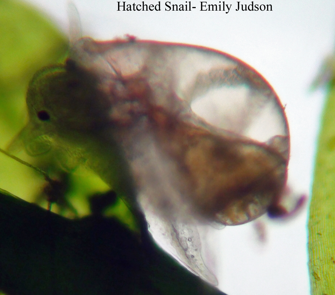
Also, the snails that have hatched since last observed, have grown significantly, and hold a very distinct shape. They also have a very unique pattern on their shell, almost swirl- like bands going around their bodies.

 This is a view of a snail from its underside.


One can observe the snail's distinct pattern on its shell. The amazing thing about the snails is how many have hatched since just last week. I could hypothesize and say that by next week, the aquarium will be full of them.


The fully grown Snail (or snail shell) remains about the same as before. It appears there is nothing inside of it, as it has stayed in the same place since last observed, and has not seemed to make any more development or progress.


Next, the Midge has grown significantly longer. Here, one can observe how much larger it has gotten. 


In addition to all of these observations, I also encountered a Cyclops! It is a pretty ordinary cyclops. It swims around very fast, has small hairs poking out from it, and two sections on the back end of it which can be determined as mobilizers for the cyclops (something to help it move).

To get these pictures and observe the things I did, I used two different microscopes with two different cameras attached to them.
The first was a microscope with an Infinity 2, which uses a self-directed light source and captures a much more magnified picture.
The second was a Laborlux 11, with a sony camera attached to capture images.


So, what I observed this week is:
- The sac of eggs are snail eggs.
- Some of the eggs have hatched and I was able to observe the snails
- The snail shell is barren
- The Midge has gotten bigger!
- A new organism, the cyclops!!

Sunday, October 21, 2012

Micro Aquarium Week One- So It Begins

On Tuesday October 16th I created a Microaquarium in my lab section. The sample I obtained came from the pond at the University of Tennessee Hospital on Cherokee Trail in Knoxville, Tennessee. This is a storm sewer sediment pond. My aquarium holds various plant species as well as micro organisms.
In my aquarium this week I found what appeared to be a sac of frog eggs. As seen in this picture, the sac is stationary and stays freely in the water. This is a gel-like round structure that contained many small cells with dots in the center of them, as shown below. 
I also found what appeared to be a midge, or what could have also been a mosquito larvae. It moves a lot, and makes many surprisingly quick movements. It is interesting to look at the lines throughout the organism and determine that it is the skeletal make up of the midge. There was only one found in my aquarium, but I am eager to see what he will look like next week!

Other than the midge and the frog eggs, I found that there wasn't much movement in my aquarium and water sample. The dirt settled at the bottom, and I did not find anything going on within it. There were, however, multiple small brown organisms swimming around, as well as little clear ones with what appeared to be green splotches in the middle of them. These all were so fast that I could not observe much more than that about them, except that there were two clear ones with green centers (which I figured was due to the presence of chlorophyll), and about four brown. 
The pond contained full sun exposure and I believe that may be responsible for most of the movement in my sample. 

What I observed this week:
- Sac of eggs (possibly frog eggs?)
- Midge/ mosquito larvae 
- Not much movement except for organisms I could not view as they moved too fast.